
•Fourteen consecutive patients with colorectal cancer were included. The following features were identified and then compared between normal and neoplastic mucosa on p-CLE images: vessel shape (straight vs irregular) vessel diameter the‘branching patterns’vessel permeability (fluorescein leakage) and blood flow (normal vs defective flux). Immunohistochemistry was used to confirm the presence and to study the morphology of vascular structures (CD-34 staining) and‘neo-vessels’(WT-1 staining) on tumour and normal mucosal sections.
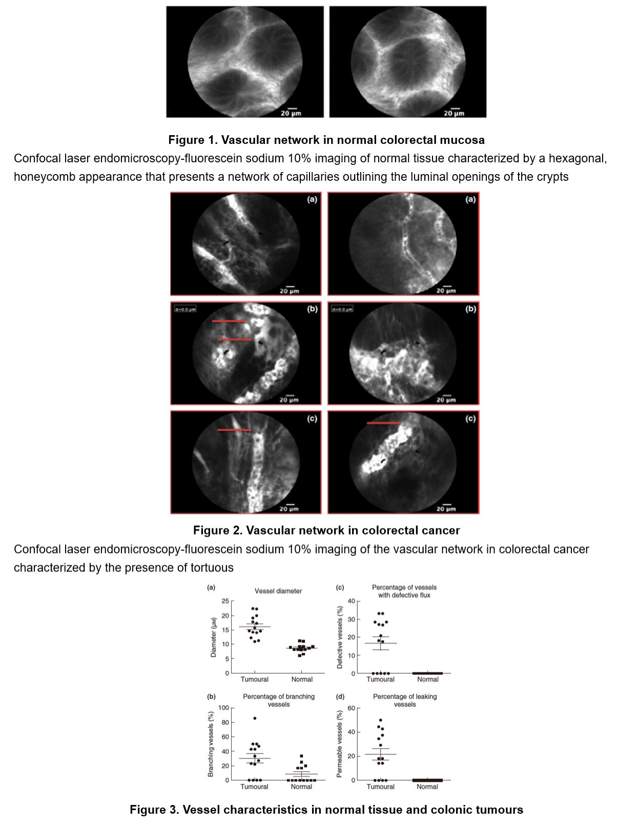
•Tumour vessels appeared as irregular, ectatic and with a highly variable calibre and branching patterns on p-CLE images. The mean diameter of tumour vessels was significantly larger than those in normal mucosa (weighted mean difference 3.38, 95% CI 2.65–4.11, P = 0.01). Similarly, ‘vessel branching’ (OR 2.74, 95% CI 1.23–6.14, P = 0.01), fluorescent dye ‘extravasation’ (OR 3.46, 95% CI 1.39–8.57, P = 0.01) were significantly more frequent in colorectal cancer than in normal colorectal mucosa. Immunohistochemistry corroborated the p-CLE findings, showing higher vascularity in tumour sections due to neoformed vessels, presenting irregular patterns.