
•Patients with suspected peripheral lung cancer based on (positron emission tomography-)CT scan underwent radial endobronchial ultrasound (rEBUS) and fluoroscopy-guided flexible bronchoscopy. After rEBUS lesion detection, an 18G needle loaded with the CLE probe was inserted in the selected airway under fluoroscopic guidance. The nCLE videos were obtained at the needle tip, followed by aspirates and biopsies. The nCLE videos were reviewed and compared with the cytopathology of the corresponding puncture and final diagnosis. Five blinded raters validated nCLE videos of lung tumours and airway/lung parenchyma twice.
•The nCLE imaging was performed in 26 patients. No adverse events occurred. In 24 patients (92%) good to high quality videos were obtained (final diagnosis; lung cancer n=23 and organising pneumonia n=1).
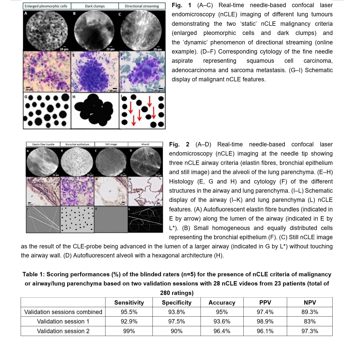
•The nCLE imaging detected malignancy in 22 out of 23 patients with lung cancer. Blinded raters differentiated nCLE videos of malignancy from airway/lung parenchyma (280 ratings) with a 95% accuracy.
•The inter-observer agreement was substantial (κ=0.78, 95% CI 0.70 to 0.86) and intra-observer reliability excellent (mean±SD κ=0.81±0.05).