
ACADEMIC HISKY
Probe-Based Confocal Laser Endomicroscopy for Diagnosis of Nasopharyngeal Carcinoma In Vivo
Lingjie Wu, ENT Institute and Otorhinolaryngology Department, Affiliated Eye and ENT Hospital, State Key Laboratory of Medical Neurobiology, Fudan University, Shanghai, China.
Objective
•This study aimed to evaluate the feasibility of using pCLE in the diagnosis of nasopharyngeal carcinoma (NPC).
Methods
•A total of 21 patients (male = 17, female = 4) with nasopharyngeal lesions were enrolled in the study.
•Biopsy specimens were collected at the imaged sites followed by a histopathological diagnosis by the pathologists, which was used as the gold standard. The pCLE images were compared to histopathological diagnosis of visualized sites.
Figures & Tables
Results
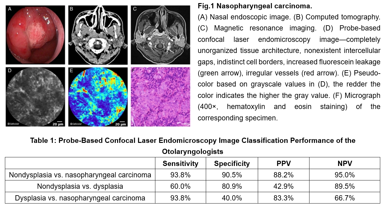
•The overall sensitivity, specificity, PPV, and NPV for diagnosis of carcinoma versus nondysplasia were 93.8%, 90.5% , 88.2% , and 95.0%.
•The sensitivity, specificity, PPV, and NPV for diagnosis of dysplasia versus nondysplasia were 60.0%, 80.9% , 42.9% , and 89.5%.
•The sensitivity, specificity, PPV, and NPV for diagnosis of carcinoma versus dysplasia were 93.8%, 40.0% , 83.3% , and 66.7%.
Conclusion
•CLE is a suitable and valid method for otolaryngologists to diagnose of NPC in vivo.
If you need more academic materials, please contact us.