
•A prospective, single-institution, nonrandomized study was conducted. No sample size calculation was performed for this observational trial. The primary objective was the description of histological rendering of normal and pathological tissues through pCLE.
•Eleven patients with hyperparathyroidism and thyroid conditions were included. A total of 104 videos showing thyroid, parathyroid, adipose tissue, muscle, laryngeal nerve, and lymph nodes were recorded. Videos were compared with visual information and pathological samples (when sampling was indicated).
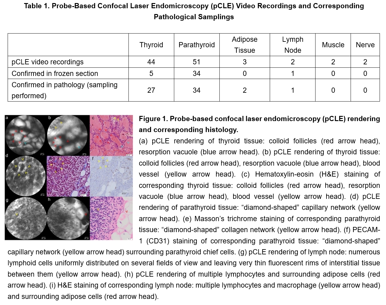
•Thyroid tissue could be identified based on the presence of colloid follicles (intensely fluorescent area surrounded by a small ridge of low-fluorescence epithelial cells) including the pathognomonic aspect of resorption vacuole. Parathyroid tissue could be identified based on a regular, "diamond-shaped" capillary network encompassing parathyroid chief cells.
•Blinded reinterpretation of pCLE videos demonstrated an 89.3% sensitivity and a 90% specificity as compared with histology in tissue recognition.