
•Freshly excised tumour samples and adjacent non-diseased sections from 50 consenting patients were stained with 0.01 % acriflavine hydrochloride and imaged using pCLE.
•All discernible pCLE features were cross-examined with conventional histopathology. Following pattern recognition training, 17 histopathologists and surgeons with no pCLE experience interpreted 50 pCLE images independently whilst blinded to histopathology results. Three-hundred and fifty pCLE image mosaics were analysed.
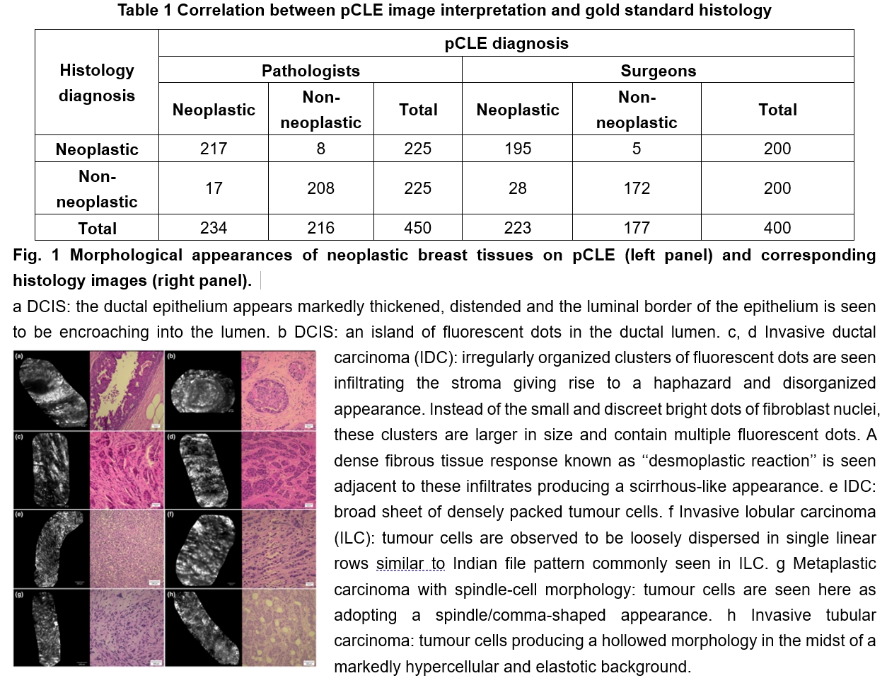
•Consistent with histopathology findings, the glandular structures, adipocytes and collagen fibres of normal breast were readily visible on pCLE images. These were distinguishable from the morphological architecture exhibited by invasive and non-invasive carcinoma.
•The mean accuracy of pCLE image interpretation for histopathologists and surgeons was 94 and 92 %, respectively.
•Overall, inter-observer agreement for histopathologists was ‘almost perfect’, k = 0.82; and ‘substantial’ for surgeons, k = 0.74.