
ACADEMIC HISKY
Analysis of 258 Different Lesions of the Central Nervous System for Real Time Histopathological Diagnosis Using Confocal Laser Endomicroscopy
Samira Daali, Department of Neurosurgery, Hospital Merheim, Cologne Medical Center, Germany.
Objective
•The aim of the current study was to evaluate CLE for a fast diagnosis of brain and spinal cord lesions in neurosurgery.
Methods
•CLE was used for an ex vivo assessment of tumor samples from 258 diverse central nervous system lesions. Additionally, traditional histology was performed on the same examined tissue as the gold standard. Nonneoplastic brain tissue served as a control.
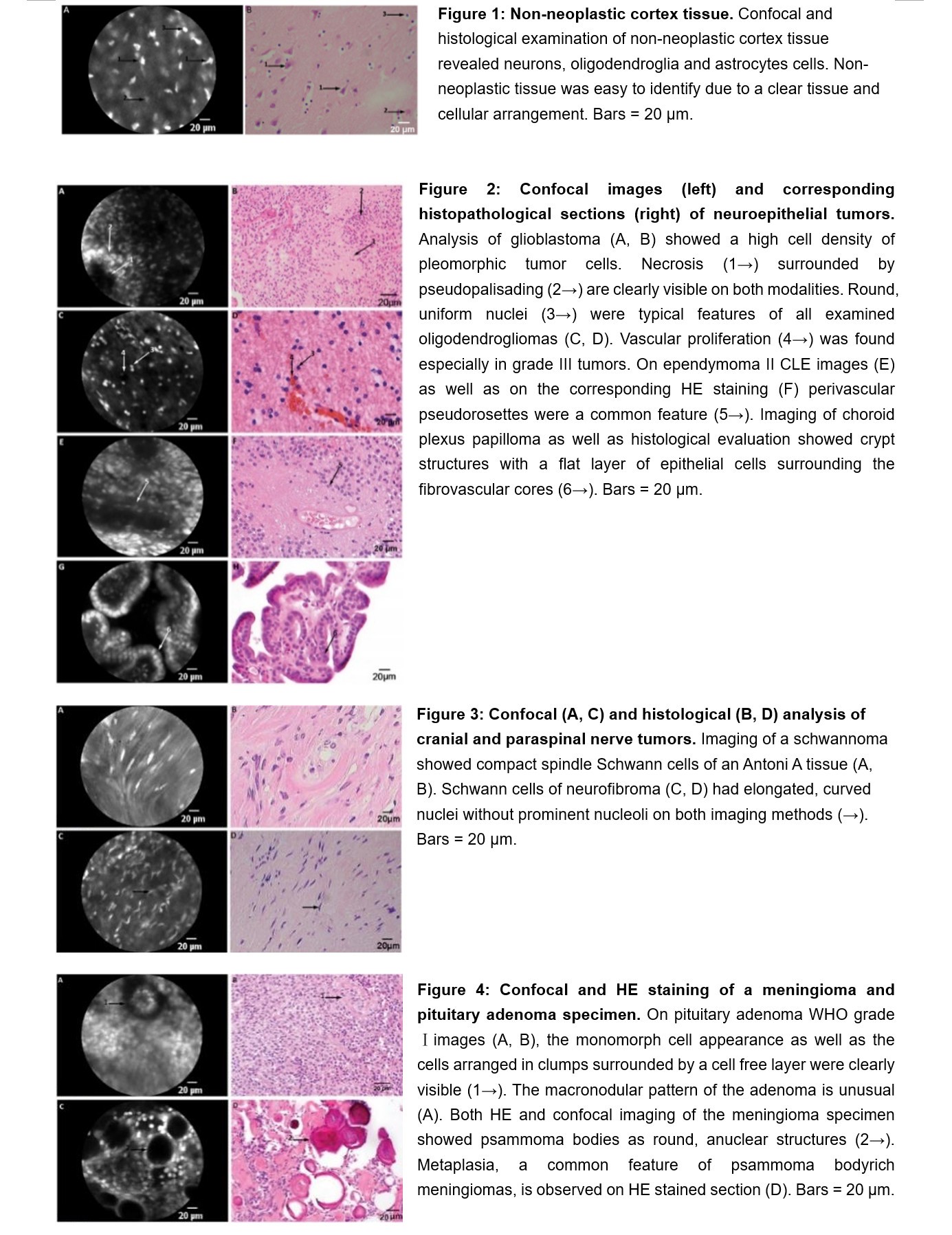
Figures & Tables
Results
•The examination of brain and spinal biopsies using CLE allowed the identification of healthy tissue, primary brain and spinal tumors, metastases, abscesses and vascular malformations with a high accuracy of 88.64%.
•Confocal imaging provided precise cellular and sub-cellular details such as psammoma bodies in meningiomas, perivascular pseudorosettes in ependymomas, microvascular proliferation in glioblastomas and mitotic activity in high-grade tumors.
Conclusion
•CLE is a promising method for distinguishing tumor from the surrounding healthy tissue, as well as for the immediate diagnosis of biopsies. Our studies have the potential to establish a faster preliminary diagnosis in an ongoing surgery as compared to current available methods. Moreover, the presented characteristic cellular and subcellular confocal features of examined tumors and non-neoplastic tissues could be used to guide future tumor surgeries to enable a more precise and safe resection.
If you need more academic materials, please contact us.