ACADEMIC HISKY
ACADEMIC HISKY
Preliminary study of confocal laser endomicroscopy for in vitro specimens of the endometrium
Jia Wen, Department of Obstetrics and Gynecology, Beijing Tsinghua Changgung Hospital, School of Clinical Medicine, Tsinghua University, China.
Objective
•This study observed and described the morphological characteristics of the endometrium of the resected uterus using confocal laser endomicroscopy.
Methods

•This prospective study included 32 patients who underwent hysterectomy.
•We used confocal laser endomicroscopy to observe the endometrium of resected uteruses and described the characteristics of endometrium in different states by comparing histopathological findings (primary objects).

Figures & Tables
Results

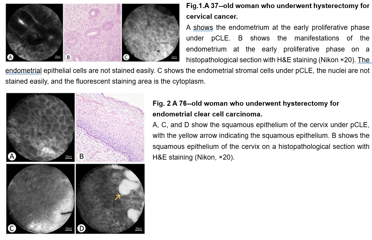
•A total of 32 patients who underwent hysterectomy for different diseases were included: 9 with endometrial carcinoma (5 with endometrioid carcinoma, 1 with endometrial serous carcinoma, 2 with clear cell carcinoma, and 1 with carcinosarcoma), 2 with atypical endometrial hyperplasia, 9 with benign diseases, 7 with cervical cancer, and 5 with ovarian cancer and borderline tumor.
•Considering histopathology as the gold standard, the diagnostic concordance rate of pCLE was 96.9% in patients with endometrial carcinoma and precancerous lesions and 100% in patients with endometrial carcinoma.

Conclusion
•Confocal laser endomicroscopy provides real-time high-resolution images of the benign endometrium and endometrial lesions. Compared with histopathology, confocal laser endomicroscopy has high diagnostic accuracy and may become an auxiliary examination tool for hysteroscopy, as it is useful for early identification of endometrial lesions, real-time diagnosis of tumor, and detection of tumor boundaries for complete tumor resection. These findings can lay a foundation for the feasible use of fertility-sparing local excision of tumor lesions by hysteroscopy.
If you need more academic materials, please contact us.
Submit